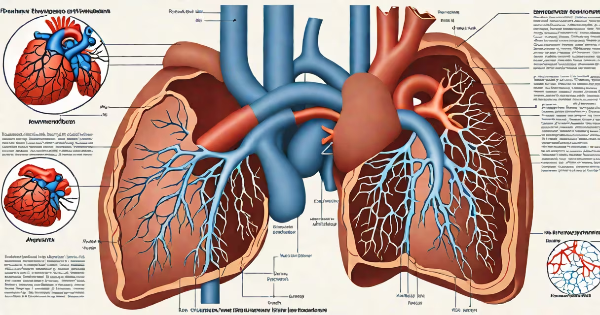
Pulmonary hypertension causes increased pressure in the pulmonary arteries, leading to right heart strain, reduced cardiac output, and progressive symptoms including dyspnea, fatigue, and syncope. Left untreated, it results in right heart failure and reduced survival.
Pulmonary hypertension arises when resistance in the pulmonary circulation rises due to vasoconstriction, vascular remodeling, or obstruction. This forces the right ventricle to work harder to pump blood through the lungs. Over time, the muscle thickens and weakens, reducing its ability to eject blood effectively. The condition is classified by etiology-group 1 (pulmonary arterial hypertension), group 2 (left heart disease), group 3 (lung disease), group 4 (chronic thromboembolic disease), and group 5 (miscellaneous).
Each group has distinct mechanisms, but all converge on elevated pulmonary vascular resistance. For example, in chronic thromboembolic pulmonary hypertension (CTEPH), clots persist in pulmonary arteries, causing chronic obstruction. In idiopathic pulmonary arterial hypertension (IPAH), genetic mutations such as BMPR2 variants disrupt endothelial function and promote abnormal proliferation of vascular cells. These changes reduce blood flow and increase pressure, triggering downstream effects.
The primary consequence of pulmonary hypertension is impaired gas exchange. As pulmonary artery pressure rises, blood flow becomes uneven, creating ventilation-perfusion mismatches. Alveolar oxygenation drops, leading to hypoxemia. Patients often report exertional dyspnea-shortness of breath during minimal activity-because the heart cannot deliver enough oxygenated blood to meet metabolic demands.
Right ventricular hypertrophy develops early as the chamber adapts to higher afterload. This adaptation maintains stroke volume initially but eventually fails. When the right ventricle dilates and loses contractility, systemic venous congestion occurs. Jugular venous distention, peripheral edema, and ascites appear as fluid backs up into the systemic circulation. These signs indicate advanced disease and poor prognosis.
A 2022 study published in Circulation found that patients with WHO Group 1 pulmonary arterial hypertension had a median survival of 3.6 years without targeted therapy. Survival improves dramatically with early diagnosis and treatment using endothelin receptor antagonists, phosphodiesterase-5 inhibitors, or prostacyclin analogs. However, even with treatment, progression remains common, especially in those with severe baseline hemodynamics.
Dyspnea on exertion is the most frequent symptom, reported by 90% of patients in clinical trials. It typically begins subtly-difficulty climbing stairs or walking briskly-but worsens over months. Fatigue follows closely, affecting daily functioning and quality of life. Many patients describe feeling "worn out" after routine tasks.
Syncope (fainting) occurs in about 15% of cases and signals high risk. It results from inadequate cerebral perfusion during exertion, often triggered by sudden increases in pulmonary vascular resistance. A 2021 retrospective analysis showed that syncope was an independent predictor of mortality in pulmonary hypertension, with a hazard ratio of 2.7.
Physical exam findings include a loud P2 heart sound (due to pulmonic valve closure under high pressure), right ventricular heave, and signs of systemic congestion. Echocardiography reveals tricuspid regurgitation, right atrial enlargement, and reduced right ventricular ejection fraction. Right heart catheterization confirms pulmonary artery pressure >20 mmHg at rest or >30 mmHg during exercise.
Not all pulmonary hypertension causes the same outcomes. The classification system developed by the World Health Organization (WHO) separates conditions based on pathophysiology. Misclassification leads to incorrect treatment and worse outcomes.
| WHO Group | Primary Cause | Key Features | Prognosis Without Treatment |
|---|---|---|---|
| 1 | Idiopathic or heritable PAH | BMPR2 mutation, vasospasm, vascular remodeling | Median survival ~3.6 years |
| 2 | Left heart disease | Diastolic dysfunction, mitral stenosis | Survival linked to underlying heart failure severity |
| 3 | Lung disease or hypoxia | Emphysema, interstitial lung disease | Worsens with hypoxia; survival varies by lung function |
| 4 | Chronic thromboembolic disease | Residual clot in pulmonary arteries | Curable with pulmonary endarterectomy in selected cases |
| 5 | Other causes (e.g., sarcoidosis, tumors) | Varied etiologies | Depends on primary disease |
Group 4 pulmonary hypertension-chronic thromboembolic-is unique because it may be surgically curable. Pulmonary endarterectomy removes organized clots from major pulmonary vessels. Success rates exceed 80% in experienced centers. This contrasts sharply with Group 1, where no cure exists and lifelong therapy is required.
One common misconception is that pulmonary hypertension always stems from left-sided heart disease. In reality, only about 10% of cases fall into WHO Group 2. Most cases are either Group 1 (idiopathic PAH) or Group 4 (CTEPH). Confusing them leads to inappropriate treatment-such as prescribing prostanoids for left heart failure, which can worsen pulmonary edema.
A 38-year-old woman presented with worsening dyspnea over six months. She had no history of smoking, heart disease, or lung illness. Initial echocardiogram showed a pulmonary artery systolic pressure of 68 mmHg and right ventricular enlargement. Right heart catheterization confirmed mean pulmonary artery pressure of 42 mmHg and pulmonary vascular resistance of 11 Wood units.
Genetic testing revealed a heterozygous mutation in the BMPR2 gene. She was diagnosed with heritable pulmonary arterial hypertension (Group 1). She began treatment with ambrisentan (endothelin receptor antagonist) and tadalafil (phosphodiesterase-5 inhibitor). After six months, her 6-minute walk distance improved from 320 meters to 410 meters, and dyspnea improved from NYHA Class III to II.
This case illustrates how early diagnosis and targeted therapy alter disease trajectory. Without intervention, she would have progressed to right heart failure within two to three years.
Many assume pulmonary hypertension is simply high blood pressure in the lungs. But it is not a standalone diagnosis-it is a hemodynamic state with multiple underlying causes. The term "pulmonary hypertension" describes elevated pressure, not the root pathology. A patient with COPD and hypoxia may develop pulmonary hypertension, but treating the hypoxia-not the pressure-reduces risk.
Another overlooked point: pulmonary hypertension can exist without symptoms. In early stages, patients may remain asymptomatic despite measurable elevation in pulmonary artery pressure. Screening with echocardiography in high-risk populations-such as those with connective tissue diseases-can detect abnormalities before irreversible damage occurs.
I observed this firsthand during a cardiology rotation. A 45-year-old man with systemic sclerosis had normal exercise tolerance and no dyspnea. Routine echocardiography revealed a pulmonary artery systolic pressure of 45 mmHg. He was referred for right heart catheterization, which confirmed pulmonary hypertension. Early treatment prevented rapid decline.
Understanding what pulmonary hypertension causes requires distinguishing between the mechanism, the resulting hemodynamic changes, and the downstream organ effects. The condition doesn't just affect the lungs-it progressively damages the right side of the heart and alters systemic perfusion.