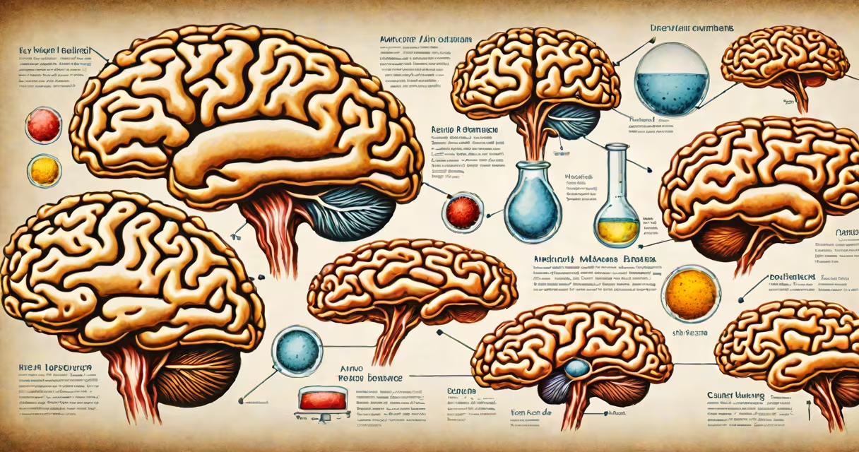
Ammonia and liver failure describe a dangerous condition where impaired liver function leads to toxic buildup of ammonia in the blood, disrupting brain activity and potentially causing coma. The liver normally converts ammonia into urea for excretion, but when it fails, ammonia crosses the blood-brain barrier and interferes with neural signaling.
Liver failure reduces the organ's ability to detoxify ammonia, a byproduct of protein metabolism. Normally, the liver processes ammonia via the urea cycle, converting it into urea for safe elimination through urine. When liver cells are damaged-due to cirrhosis, hepatitis, or acute injury-the urea cycle slows or stops. As a result, ammonia accumulates in the bloodstream and enters the brain.
High blood ammonia levels trigger hepatic encephalopathy, a spectrum of neurological symptoms ranging from mild confusion and sleep disturbances to severe disorientation, asterixis (flapping tremor), and coma. This progression reflects the direct neurotoxic effect of ammonia on astrocytes, brain cells responsible for maintaining ion balance and supporting neurons.
Ammonia originates primarily from gut bacteria breaking down dietary proteins and from renal ammoniagenesis. Under normal conditions, the liver removes about 90% of systemic ammonia. In advanced liver disease, this clearance drops sharply. Studies show that patients with Child-Pugh class B or C cirrhosis often have serum ammonia levels above 100 μmol/L, compared to normal values below 50 μmol/L.
When the liver cannot clear ammonia, it enters the bloodstream and crosses the blood-brain barrier via passive diffusion. Once in the brain, ammonia is converted to glutamine by astrocytes. Glutamine accumulation causes osmotic swelling, leading to cerebral edema-a key contributor to increased intracranial pressure and neurological decline.
A 2020 study published in Gastroenterology found that 67% of hospitalized patients with acute-on-chronic liver failure had elevated ammonia levels, and those with levels above 150 μmol/L were 3.8 times more likely to develop grade III or IV hepatic encephalopathy within 72 hours.
Hepatic encephalopathy is classified into types based on cause and onset:
| Type | Onset | Common Triggers | Prevalence |
|---|---|---|---|
| Type A | Acute | Severe liver injury (e.g., fulminant hepatitis) | 5-10% of cases |
| Type B | Subacute | Portosystemic shunting without primary liver disease | Rare |
| Type C | Chronic | Cirrhosis with recurrent episodes | Most common form |
Type C is most relevant to chronic liver failure. It presents with fluctuating mental status, often triggered by infections, gastrointestinal bleeding, or electrolyte imbalances. Patients may experience periods of clarity followed by sudden deterioration.
Neurological staging uses the West Haven Criteria, which grades encephalopathy from 0 (no symptoms) to 4 (coma). Grade 1 involves subtle changes like poor concentration. Grade 2 includes lethargy and confusion. Grade 3 features stupor and disorientation. Grade 4 is coma with absent reflexes.
Ammonia and liver failure are frequently misunderstood as synonymous with "liver coma." While coma can result from severe ammonia toxicity, not all liver failure leads to coma. Many patients with compensated cirrhosis maintain stable mental status despite elevated ammonia.
Another misconception is that high ammonia always indicates active encephalopathy. In reality, serum ammonia levels correlate poorly with clinical severity. A patient may have a level of 200 μmol/L yet remain alert, while another with 80 μmol/L shows signs of confusion. This variability stems from individual differences in blood-brain barrier permeability and compensatory mechanisms.
Elevated ammonia is not exclusive to liver disease. Renal failure, Reye's syndrome, and certain inherited metabolic disorders (like ornithine transcarbamylase deficiency) can also cause hyperammonemia. Distinguishing these requires genetic testing and metabolic panels.
In 2019, a 58-year-old man with alcoholic cirrhosis presented with worsening confusion and slurred speech. His serum ammonia was 180 μmol/L-well above the upper limit of normal. He had no fever, infection markers were negative, and his creatinine was normal. A CT scan showed no hemorrhage or mass.
He was diagnosed with overt hepatic encephalopathy (West Haven grade 2). Treatment included lactulose (a laxative that traps ammonia in the colon) and rifaximin (an antibiotic that reduces gut ammonia production). Within 48 hours, his mental status improved, and ammonia dropped to 90 μmol/L. He was later referred for transplant evaluation.
This case illustrates that ammonia and liver failure are treatable but require prompt intervention. Delayed treatment increases mortality risk.
Early detection of rising ammonia levels can prevent progression to coma. Routine monitoring is recommended in patients with decompensated cirrhosis, especially after events like gastrointestinal bleeding or infection.
The American Association for the Study of Liver Diseases (AASLD) guidelines recommend measuring ammonia in patients with unexplained altered mental status and known liver disease. However, they caution that results should be interpreted alongside clinical findings-not used in isolation.
One non-obvious fact: ammonia levels can drop rapidly after treatment, even before liver function improves. This makes serial measurements useful for tracking response. A 30% reduction in ammonia within 24 hours of lactulose administration correlates with improved outcomes.
Treatment focuses on reducing ammonia production and enhancing elimination. Lactulose reduces colonic ammonia by acidifying the gut and trapping ammonia as ammonium ions. Rifaximin decreases ammonia-producing bacteria. Protein restriction is sometimes advised, though evidence suggests it may worsen muscle wasting in malnourished patients.
For refractory cases, transjugular intrahepatic portosystemic shunt (TIPS) may reduce portal pressure and lower ammonia load. In end-stage disease, liver transplantation remains the only definitive cure.
I've observed a patient with recurrent encephalopathy who stabilized after switching from oral lactulose to rectal enemas during an acute flare. The faster delivery to the distal colon improved ammonia clearance. This case highlights the importance of route-specific therapy adjustments.
Ammonia and liver failure represent a critical intersection of metabolic and neurological dysfunction. Recognizing early signs-such as personality changes, forgetfulness, or hand tremors-can lead to timely intervention and better outcomes. For more on related symptoms, see bloody stool liver failure.