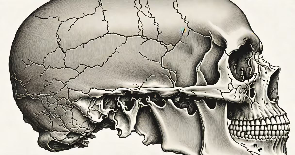
The skull of the human body is a rigid bony structure composed of 22 bones that encase and protect the brain while forming the structure of the face. It consists of the neurocranium, which surrounds the brain, and the viscerocranium, which supports the facial features. This structure provides attachment points for muscles involved in chewing, facial expression, and jaw movement.
The skull's primary function is protection. The brain occupies about 1,300 cm³ of space, and the skull maintains a stable environment within a rigid, non-expandable container. A fracture in the skull can lead to direct injury to the brain tissue, cerebrospinal fluid leakage, or intracranial hemorrhage. According to the Centers for Disease Control and Prevention (CDC), traumatic brain injuries account for over 2 million emergency department visits annually in the U.S., many involving skull trauma.
Each bone in the skull serves a specific role. The frontal bone forms the forehead and upper part of the eye sockets. The parietal bones make up the sides and roof of the cranium. The temporal bones house the inner ear structures and form part of the jaw joint. The occipital bone contains the foramen magnum, through which the spinal cord connects to the brainstem. The sphenoid and ethmoid bones contribute to the base of the skull and support the nasal cavity and sinuses.
The skull functions as both a protective casing and a structural scaffold. Its bones are fused together at joints called sutures, which allow minimal movement during development but become immobile in adulthood. These sutures-such as the coronal, sagittal, and lambdoid sutures-help absorb impact and distribute forces across the skull during trauma.
When a blow to the head occurs, the skull's rigidity prevents deformation, but this also means force is transmitted directly to the brain. A concussion often results from rapid acceleration or deceleration of the head, causing shearing of neural tissues even without visible skull fracture. The skull's shape helps minimize rotational stress in some directions, but high-velocity impacts can still cause complex internal injuries.
For example, a fall from a height resulting in a 6-meter drop leads to a calculated impact velocity of approximately 10.8 m/s. At this speed, the skull may withstand compression forces up to 1,500 newtons before fracturing, depending on bone density and age. Studies show that skull fractures occur in about 10% of severe head injuries, with the most common sites being the temporal bone and the orbital rim.
The skull divides into two functional regions: the neurocranium and the viscerocranium. The neurocranium includes eight bones-the frontal, two parietals, two temporals, occipital, sphenoid, and ethmoid-that enclose the cranial cavity. The viscerocranium comprises 14 bones responsible for facial structure: two maxillae, two zygomatics, two palatines, two inferior nasal conchae, two lacrimals, the vomer, and two mandibles.
| Bone Group | Number of Bones | Primary Functions |
|---|---|---|
| Neurocranium | 8 | Protects brain; forms cranial cavity; houses sensory organs |
| Viscerocranium | 14 | Supports face; enables mastication and facial expression; forms nasal passages |
Some bones, like the mandible, are mobile. The mandible articulates with the temporal bones via the temporomandibular joints (TMJ), allowing jaw movement for speaking and eating. The TMJ is unique among skull joints because it permits both rotational and translational motion. Dislocation or dysfunction here affects speech clarity and chewing efficiency.
A lesser-known fact: the skull contains air-filled spaces called sinuses. The frontal, ethmoid, sphenoid, and maxillary sinuses reduce bone weight and humidify inhaled air. They also contribute to voice resonance. Infections in these cavities-like sinusitis-affect up to 15% of adults annually, according to the American Academy of Otolaryngology-Head and Neck Surgery.
People often assume the skull is a single solid bone. In reality, it's a composite of 22 separate bones joined by sutures. These sutures remain flexible during infancy to accommodate brain growth and close gradually between ages 18 and 25. Premature closure of sutures-known as craniosynostosis-can restrict brain development and requires surgical correction.
Another misconception is that skull fractures always involve visible external wounds. In reality, depressed skull fractures may appear subtle or absent externally. A CT scan detects these injuries in 90% of cases, whereas plain X-rays miss up to 40% due to overlapping bone structures.
Some confuse the skull with the spine or neck vertebrae. While the skull connects to the cervical spine at the atlanto-occipital joint, the spine extends downward and supports the trunk. The first cervical vertebra (C1), or atlas, holds the skull's weight and allows nodding motion. Without proper alignment between the skull and spine, conditions like atlantoaxial instability can develop, leading to neurological deficits.
In forensic science, the skull is critical for identifying remains. Facial reconstruction using cranial morphology and software like FaceLab can estimate appearance with 85% accuracy in known cases. Anthropologists use skull measurements-such as cranial index (width/length ratio)-to classify human populations and trace migration patterns.
I once observed a cadaver dissection where a student misidentified the zygomatic arch as part of the temporal bone. Correct identification required palpating the zygomatic process of the temporal bone and comparing it to the zygomatic bone's lateral projection. This mistake highlights how spatial understanding of the skull is essential in medical training.
Even parts of the lower leg, like the calf, depend on the skull's sensory feedback loop. Proprioception from the head and neck influences posture and gait. When skull alignment is off, it can trigger compensatory movements in the lower limbs, affecting walking patterns and increasing injury risk.
The skull of the human body is not merely a passive shield-it is a changing, integrated structure that shapes perception, movement, and survival. Its complexity demands precision in diagnosis, treatment, and education. Understanding its anatomy is essential for anyone working in medicine, forensics, or rehabilitation.