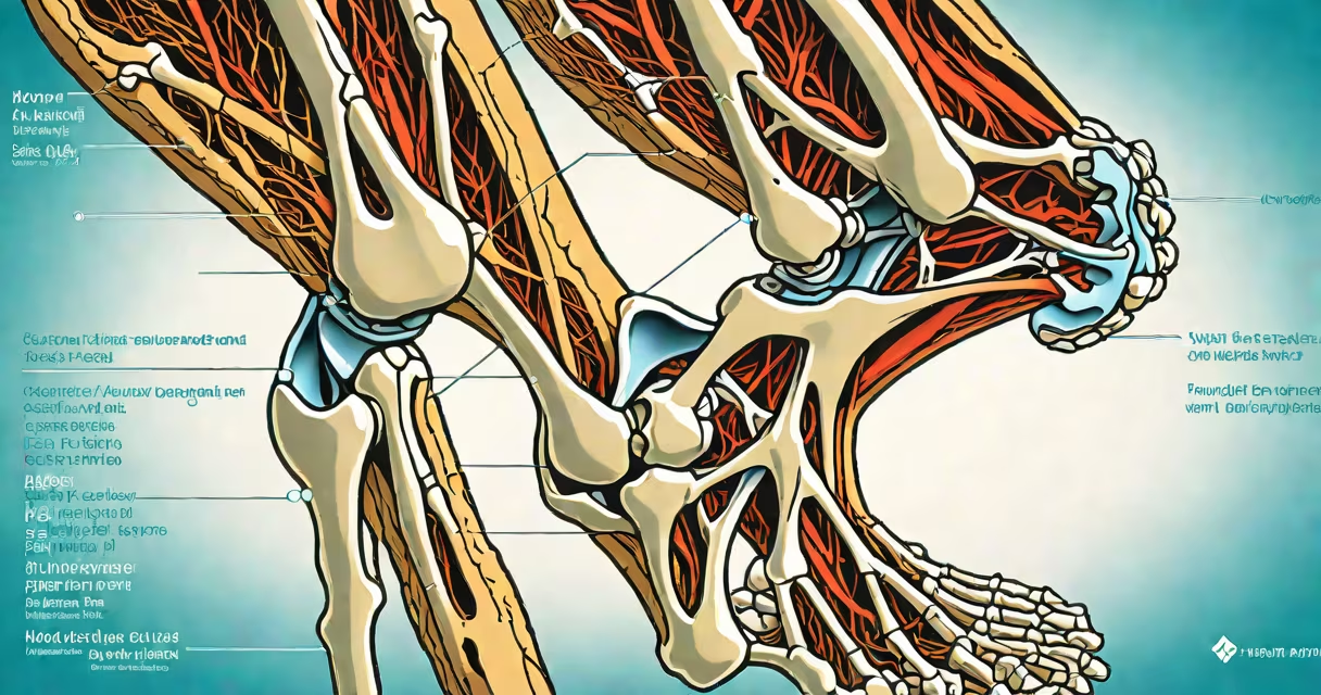
The anterior cruciate ligament (ACL) is a critical stabilizing structure located in the center of the knee joint, running diagonally between the femur (thigh bone) and the tibia (shin bone). It prevents excessive forward movement of the tibia relative to the femur and provides rotational stability during changing motion.
Specifically, the ACL originates from the medial aspect of the lateral femoral condyle and inserts into the intercondylar notch of the tibia, near the center of the knee's weight-bearing surface. This central position allows it to act as a primary restraint against anterior translation and internal rotation of the tibia during activities like cutting, jumping, or sudden deceleration.
According to the American Academy of Orthopaedic Surgeons (AAOS), the ACL measures approximately 3 cm in length and 1 cm in width in adults, making it one of the most frequently injured ligaments in the knee. Its location deep within the joint capsule makes it vulnerable to trauma during sports involving rapid changes in direction, such as soccer, basketball, and skiing.
The ACL does not work in isolation. During walking, running, or pivoting, it engages alongside other knee structures-including the posterior cruciate ligament (PCL), medial and lateral collateral ligaments, and the menisci-to maintain joint integrity. When the knee is bent at 90 degrees, the ACL becomes taut, limiting forward displacement of the tibia.
A common scenario leading to ACL injury involves a non-contact mechanism: planting the foot while changing direction, resulting in a sudden inward twist of the knee. The ACL resists this shear force, but if the load exceeds its tensile strength-approximately 2,000 newtons-it can rupture. This typically occurs during high-speed maneuvers where the athlete cannot control the knee's rotation.
For example, a soccer player attempting a sharp cut to avoid a defender may experience a loud pop in the knee, followed by immediate swelling and instability. Imaging confirms the tear, usually seen on MRI as a discontinuity in the ligament's normal linear appearance.
ACL injuries are classified by severity: grade I (mild sprain), grade II (partial tear), and grade III (complete rupture). A complete tear-common in athletes-results in significant joint instability and often requires surgical reconstruction.
One misconception is that ACL injuries always result from direct contact. In reality, 70% of ACL tears occur without collision, according to research published in the Journal of Orthopaedic Research. Another is that recovery takes months. While rehabilitation typically lasts 6-9 months, return to sport is not guaranteed until neuromuscular control and strength metrics meet criteria set by physical therapists.
The ACL is sometimes confused with the PCL, which lies behind it. The PCL prevents backward tibial displacement and is less commonly injured. Unlike the ACL, which is prone to non-contact trauma, PCL injuries often stem from dashboard impacts in car accidents or falls onto a bent knee.
| Structure | Location Relative to ACL | Primary Function |
|---|---|---|
| Posterior Cruciate Ligament (PCL) | Behind the ACL | Resists posterior tibial translation |
| Medial Collateral Ligament (MCL) | On the inner side of the knee | Stabilizes against valgus stress |
| Lateral Collateral Ligament (LCL) | On the outer side of the knee | Resists varus stress |
| Menisci | Between femur and tibia, lateral and medial | Shock absorption and load distribution |
These structures form a network of support. Damage to any one can affect ACL function, but the ACL's central role makes its injury particularly disabling for athletic performance.
In 2022, a Division I college football player tore his ACL during a game while attempting to evade a tackle. He landed on his planted leg with his knee slightly bent and rotated inward. The injury was diagnosed via MRI, which showed a complete disruption of the ACL fibers at the femoral attachment site.
Surgical reconstruction using a hamstring autograft was performed six weeks later. Post-op rehabilitation included progressive strengthening, balance training, and sport-specific drills. After nine months, he returned to play, though his performance metrics-such as sprint speed and change-of-direction time-remained below pre-injury levels.
This case illustrates that even with successful surgery, long-term outcomes vary. A study in Arthroscopy found that 60% of athletes return to their prior level of competition, but only 40% achieve full functional recovery at two years post-surgery.
Many people assume the ACL is visible during knee surgery because it lies deep inside the joint. However, surgeons access it through arthroscopic portals, not open incisions. The ACL is not palpable during physical exams-its integrity must be confirmed via imaging or clinical tests like the Lachman test or anterior drawer test.
Another error is equating ACL pain with arthritis. While both conditions cause knee discomfort, ACL tears typically present with acute swelling, a sensation of the knee "giving way," and inability to continue activity. Arthritis develops gradually and causes stiffness, especially in the morning.
There is also confusion between ACL location and the site of tendon grafts used in reconstruction. Grafts are harvested from the patellar tendon, hamstring tendons, or quadriceps tendon-structures separate from the ACL itself-but they are placed in the same anatomical space to restore function.
I observed an ACL reconstruction procedure in a hospital operating room. The surgeon used a camera inserted through a small incision to visualize the joint. The ACL was completely torn, with frayed ends and a gap in the femoral tunnel. A hamstring graft was prepared and passed through drill holes drilled into the femur and tibia, then secured with screws. The entire process took about 90 minutes under general anesthesia.
After the surgery, the patient reported immediate relief from instability, though full recovery required months of therapy. The procedure restored mechanical stability, but neurological retraining was necessary to regain confidence in movement.
Knowing where your ACL is located helps clarify why certain movements are risky and why proper conditioning reduces injury risk. The ACL is not just a band-it's a biomechanical anchor that enables complex motion, and its health is essential for mobility and athletic function.