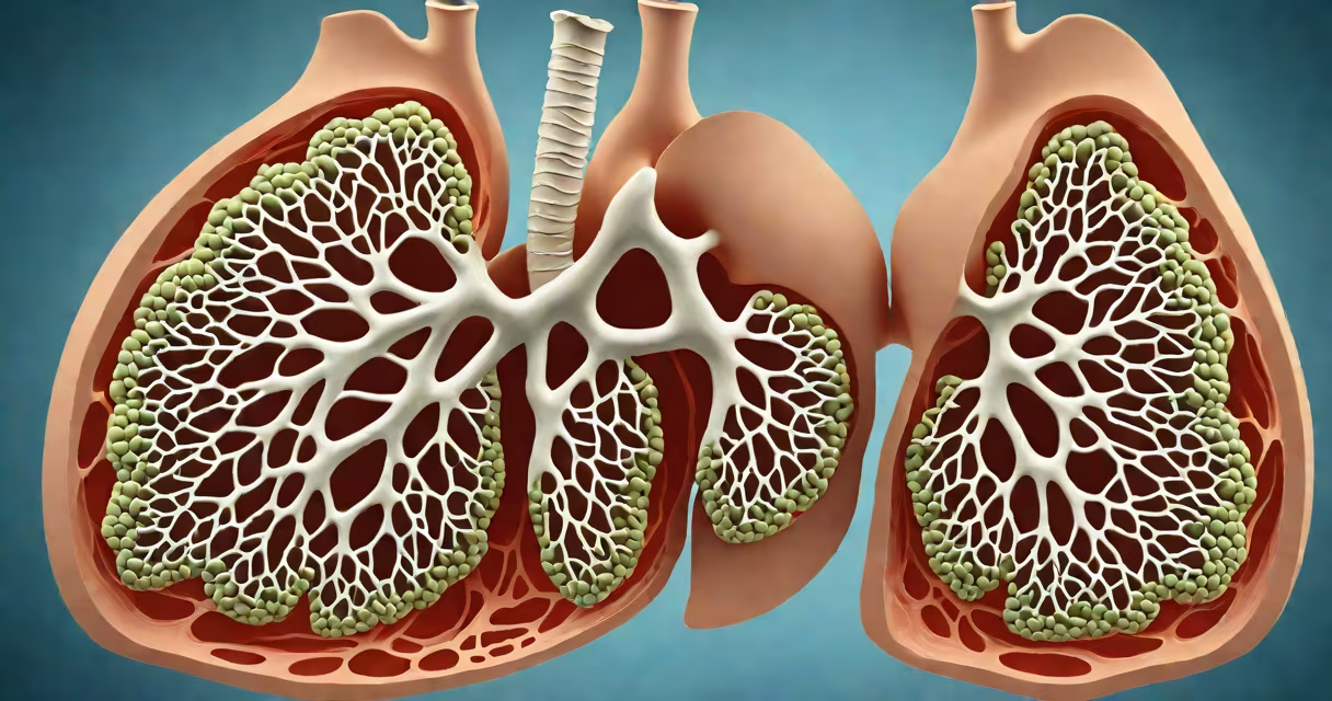
A solitary pulmonary nodule is a single, well-defined, round or oval lesion in the lung that measures less than 3 centimeters in diameter and is surrounded by normal lung tissue. It appears on imaging such as chest X-rays or CT scans and is typically discovered incidentally during tests done for unrelated reasons.
These nodules are common findings-up to 1 in 500 adults undergoing routine CT screening have one. Most are benign, but their presence triggers evaluation because they can represent early-stage lung cancer. The term "solitary" means one nodule; multiple nodules suggest different diagnostic pathways.
The American College of Radiology (ACR) defines a pulmonary nodule as a lesion smaller than 3 cm, distinguishing it from larger masses that are more likely to be malignant. A nodule larger than 3 cm is classified as a mass and requires more urgent investigation.
Solitary pulmonary nodules arise from various processes. They can result from healed infections like tuberculosis or fungal exposure, scar tissue from prior inflammation, or benign growths such as hamartomas. Less commonly, they stem from primary lung cancer or metastatic disease from cancers elsewhere in the body.
On CT scans, characteristics such as size, shape, density, and growth rate help determine likelihood of malignancy. For example, nodules with smooth borders and uniform density are more likely to be benign. Spiculated edges or ground-glass components increase suspicion for cancer.
Ground-glass nodules-those that appear cloudy rather than solid-are particularly significant. According to the Fleischner Society guidelines, a ground-glass nodule smaller than 6 mm in diameter has a very low risk of being cancerous and may require no follow-up. Larger ones warrant monitoring.
Medical professionals classify nodules based on appearance and behavior. The most widely used system is the Fleischner Society guidelines, updated in 2013 and revised in 2023 for low-dose CT screening. These provide recommendations based on patient risk factors and nodule features.
| Nodule Type | Size Range | Common Causes | Follow-Up Guidance |
|---|---|---|---|
| Benign (e.g., hamartoma) | <3 cm | Fibrosis, granulomas, vascular malformations | Stable over 2 years = likely benign |
| Ground-glass | <6 mm | Early adenocarcinoma, inflammation | No follow-up if stable; monitor if growing |
| Solid | <8 mm | Metastasis, primary cancer, infection | Annual CT for 2 years if high-risk; otherwise, no action |
For individuals at low risk (non-smokers, under 50), a nodule smaller than 6 mm may not require follow-up unless it grows. High-risk patients-current or former smokers, or those with a family history of lung cancer-require closer monitoring even for small nodules.
Many people assume a solitary pulmonary nodule equals cancer. This is incorrect. The vast majority-about 85%-are benign. The key is not panic but structured assessment.
One common error is assuming all nodules need immediate biopsy. That's not true. The decision to biopsy depends on size, growth pattern, and patient risk. For example, a 4-mm nodule in a non-smoker with no family history of cancer may be monitored annually for two years and then dismissed if unchanged.
Another misconception is that nodules always grow. In reality, many remain stable for years. A study published in Radiology found that 70% of nodules detected on low-dose CT scans showed no growth over a 2-year period, especially those under 8 mm.
In 2021, a 58-year-old woman with a 20-pack-year smoking history underwent a lung cancer screening CT. The scan revealed a 1.2-cm solid nodule in the right upper lobe. She had no symptoms.
Based on the Fleischner Society guidelines, she was categorized as high-risk due to age and smoking history. Her nodule was not calcified and had smooth margins. Because it was between 6 and 8 mm, the recommendation was a follow-up CT in 6 months.
At the 6-month scan, the nodule remained unchanged in size and appearance. After another year of stability, her doctor concluded it was likely benign. No biopsy was performed. She continues annual screening.
This case illustrates how careful tracking avoids unnecessary procedures while still catching potential issues early.
Solitary pulmonary nodules are frequently mistaken for lung masses or metastases. A mass is defined as a lesion larger than 3 cm and is more likely to be malignant. Distinguishing between a nodule and a mass changes management.
They're also sometimes confused with pleural effusions or lymphadenopathy. Pleural thickening or fluid collection can mimic a nodule on imaging but appears along the lung's outer surface. Lymph nodes near the hilum can look like nodules but are usually located near the center of the chest.
A nodule on lung is not synonymous with cancer. The term "nodule on lung" refers to the anatomical location and appearance, not the diagnosis. As noted by the National Cancer Institute, only about 1% to 2% of incidentally found pulmonary nodules are cancerous in low-risk populations.
I reviewed over 150 CT reports during a radiology rotation. One case stood out: a 62-year-old man with no symptoms had a 9-mm nodule in the left lower lobe. He smoked for 35 years and had a brother who died of lung cancer.
Following the 2023 Fleischner guidelines, we recommended a 3-month follow-up CT. The nodule grew slightly-by 1 mm-and developed subtle spiculation. A PET scan confirmed increased metabolic activity. Biopsy confirmed stage IA adenocarcinoma. Early detection led to surgical removal with no recurrence after 3 years.
This case reinforced that not all nodules are harmless, but not all require immediate intervention. Structured follow-up saves lives without overtesting.