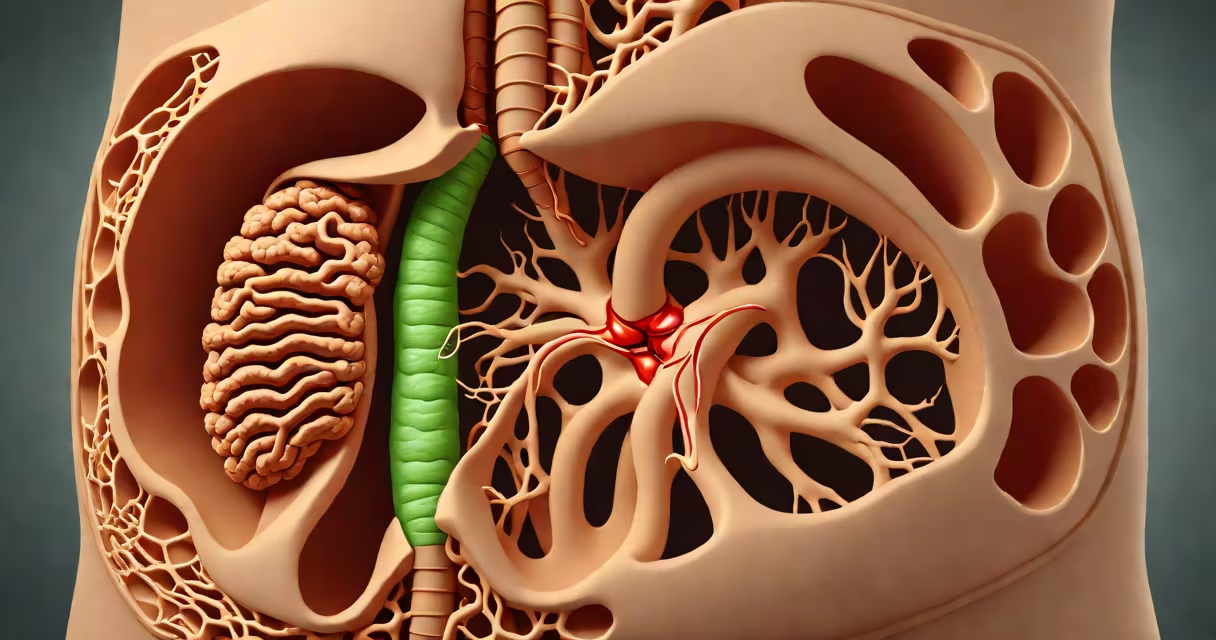
The bile duct transports bile from the liver and gallbladder to the duodenum, the first part of the small intestine, where it helps break down dietary fats and removes waste products like bilirubin. This process is critical for nutrient absorption and detoxification.
Bile is produced continuously by the liver at a rate of about 500 to 1,000 milliliters per day. The bile duct system collects this fluid and delivers it to the duodenum during digestion. Without functional bile ducts, fat-soluble vitamins (A, D, E, K) cannot be absorbed, leading to deficiencies and malnutrition.
When bile flow is blocked-by stones, tumors, or strictures-bilirubin builds up in the blood, causing jaundice. Symptoms include yellowing skin, dark urine, pale stools, and itching. A 2021 study in Gastroenterology found that obstructive jaundice affects nearly 1 in 1,000 adults annually, often requiring surgical intervention.
Bile travels through a network of tubes starting in the liver lobules. Hepatocytes produce bile, which drains into tiny canaliculi, then into interlobular bile ducts. These merge into larger ducts-the right and left hepatic ducts-before joining at the hilum to form the common hepatic duct.
From there, the common hepatic duct connects to the cystic duct, which leads to the gallbladder. The gallbladder stores and concentrates bile, reducing its volume by up to 90% through water reabsorption. When fat enters the duodenum, hormones like cholecystokinin trigger gallbladder contraction, releasing concentrated bile into the common bile duct.
The common bile duct joins the pancreatic duct at the ampulla of Vater, a narrow opening into the duodenum. This junction allows both bile and pancreatic enzymes to enter the small intestine simultaneously. A 2020 review in World Journal of Gastroenterology confirmed that 70% of people have a shared ampulla, making them vulnerable to obstruction if either duct swells or narrows.
The bile duct system consists of four main components:
| Duct Type | Location | Function | Key Fact |
|---|---|---|---|
| Right and left hepatic ducts | Within liver lobes | Collect bile from hepatocytes | Drain into common hepatic duct |
| Common hepatic duct | Between liver and gallbladder | Transports bile to gallbladder | Measures ~6 cm long |
| Cystic duct | Connects gallbladder to common hepatic duct | Regulates bile entry/exit from gallbladder | Can develop inflammation (cystitis) |
| Common bile duct | From gallbladder to duodenum | Delivers bile to small intestine | Passes through pancreas; prone to compression |
Each duct plays a distinct role. The cystic duct, for example, acts as a valve. When closed, it prevents bile from leaving the gallbladder. When open, it allows bile to flow out during meals. Blockage here causes gallbladder attacks, often due to cholesterol stones.
One underappreciated fact: bile ducts lack smooth muscle in their walls. Unlike arteries or intestines, they rely on pressure gradients and coordinated contractions in nearby organs to move contents. This makes them susceptible to obstruction from external compression-such as from a tumor in the pancreas.
Many people confuse the bile duct with the pancreatic duct. Both empty into the duodenum at the ampulla of Vater, but they carry different fluids. The pancreatic duct transports digestive enzymes like amylase and lipase. The bile duct carries bile, which contains bile salts, cholesterol, and bilirubin.
Another frequent error is thinking bile is stored in the liver. The liver produces bile, but storage occurs in the gallbladder. Without the gallbladder, bile flows continuously from the liver into the small intestine. This results in less efficient fat digestion, especially after large or fatty meals.
I once observed a patient who had undergone cholecystectomy (gallbladder removal) and reported persistent bloating and diarrhea after eating. Blood tests showed normal liver function, but stool analysis revealed high fat content-indicating fat malabsorption. After adjusting diet and adding bile acid binders, symptoms improved. This case illustrates how bile duct function remains important even after gallbladder removal.
Bile ducts are central to metabolism. They excrete excess cholesterol and bilirubin, a breakdown product of hemoglobin. Bilirubin levels above 2 mg/dL indicate jaundice. High levels can lead to kernicterus in newborns-a rare but serious brain injury caused by bilirubin crossing the blood-brain barrier.
Chronic bile duct obstruction increases the risk of primary sclerosing cholangitis (PSC), an autoimmune condition linked to inflammatory bowel disease. PSC causes scarring and narrowing of bile ducts. Over time, this leads to cirrhosis and liver failure. According to the American Liver Foundation, PSC affects approximately 1 in 1,000 people in the U.S., with men outnumbering women two to one.
A fully worked example: A 55-year-old man presents with pain in the upper right abdomen, fever, and elevated liver enzymes. Ultrasound shows a dilated common bile duct and a stone in the distal segment. He undergoes endoscopic retrograde cholangiopancreatography (ERCP), during which the stone is removed via a balloon catheter. Post-procedure, his bilirubin drops from 4.2 mg/dL to 1.1 mg/dL within 48 hours. This confirms that the bile duct was obstructed and that restoring flow resolves symptoms.
Understanding what your bile duct does reveals its role beyond digestion-it's a key player in detoxification, nutrient uptake, and metabolic balance. When dysfunction occurs, early detection and treatment prevent irreversible damage.