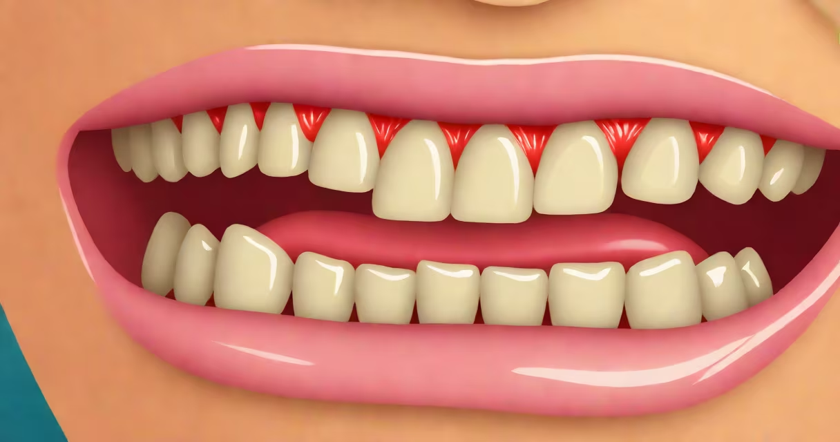
Leukoplakia is a condition marked by thickened, white patches on the mucous membranes inside the mouth, most commonly caused by prolonged irritation from tobacco use, ill-fitting dentures, or chronic friction. It affects about 1 in 1,000 adults globally and carries a 3% to 5% risk of progressing to oral squamous cell carcinoma over time.
The lesions form when epithelial cells in the mouth undergo hyperkeratosis-a buildup of keratin-as a response to persistent stress. These patches do not rub off with a cotton swab, unlike fungal infections such as thrush. They typically appear on the gums, inner cheeks, or under the tongue and may feel rough or lumpy to the touch.
According to the American Dental Association, leukoplakia is diagnosed through visual inspection and confirmed via biopsy. The presence of abnormal cells in the tissue sample determines whether the lesion is benign, dysplastic, or malignant. Early detection reduces the chance of progression to cancer.
Tobacco use remains the leading cause of leukoplakia. Smokers have a 6.5 times higher risk of developing the condition compared to non-smokers, based on data from the National Institute of Dental and Craniofacial Research (NIDCR).
Other mechanical irritants include sharp teeth, poorly fitted dental appliances, or habitual cheek biting. A 2021 study in the Journal of Oral Pathology & Medicine found that 38% of patients with leukoplakia had a history of trauma from dental hardware.
Chronic irritation from hot foods, alcohol consumption, or spicy diets contributes less frequently but still plays a role. Human papillomavirus (HPV) infection is a lesser-known factor-HPV-16 has been detected in 12% of leukoplakia cases, particularly in younger patients without tobacco exposure.
| Common Cause | Prevalence in Cases | Key Risk Factor |
|---|---|---|
| Tobacco use (smoking/chewing) | 70-80% | Duration and frequency of use |
| Dental appliance irritation | 15-20% | Poor fit or sharp edges |
| Chronic friction (cheek biting) | 5-10% | Repetitive trauma |
| HPV infection (especially HPV-16) | 10-15% | Age under 40, no tobacco history |
Leukoplakia is often mistaken for candidiasis (thrush), which also presents as white patches. However, thrush can be wiped away with a gauze pad, while leukoplakia cannot. Misdiagnosis occurs in up to 12% of primary care settings, according to a 2020 review in the British Journal of General Practice.
Another common error is assuming all white patches are harmless. Some leukoplakia lesions show dysplasia-abnormal cell growth-on histology. A 2019 study in the International Journal of Cancer reported that 17% of biopsied leukoplakia cases showed high-grade dysplasia, a precursor to cancer.
Patients sometimes delay seeking care because they assume the patch is temporary or painless. But even asymptomatic lesions require monitoring. The absence of pain does not indicate safety.
A clinician begins with a visual exam and tactile assessment. If the patch is suspicious-irregular borders, redness, ulceration-it is referred for biopsy. The gold standard is a punch biopsy, where a small sample of tissue is removed and examined under a microscope.
For example, a 54-year-old man presented with a 1.2 cm white patch on his left buccal mucosa. He smoked two packs a day for 30 years. The lesion did not resolve after two weeks of stopping tobacco. A biopsy revealed moderate dysplasia. He underwent surgical excision and has since remained cancer-free after five years of follow-up.
After diagnosis, treatment focuses on removing the irritant. Tobacco cessation is mandatory. Dentures are adjusted or replaced. Patients are monitored every six months with clinical exams and, if needed, repeat biopsies.
Topical treatments like 5% 5-fluorouracil cream have shown limited success in reducing lesion size but are not curative. Surgical removal is recommended for lesions with dysplasia or those that persist after six months of irritant removal.
Leukoplakia is not always linked to tobacco. While smoking is the biggest risk factor, a subset of cases-especially in non-smokers aged 30-50-arises from autoimmune conditions like lichen planus, which mimics leukoplakia clinically but differs histologically.
Another misconception is that all leukoplakia leads to cancer. Only a minority progress. The NIDCR reports that approximately 3% of untreated cases develop into invasive cancer within ten years. However, recurrence rates are high-up to 40% in patients who continue tobacco use.
Leukoplakia is not contagious. It does not spread through saliva or kissing. This distinction is important for patient education and mental health support.
I once observed a patient with a large, irregular patch on the floor of the mouth that had persisted for over a year. Despite multiple visits, he avoided biopsy due to fear. When he finally consented, the pathology report confirmed early-stage squamous cell carcinoma. Early intervention prevented metastasis.
Leukoplakia is not a single disease but a clinical sign of underlying cellular stress. Its causes are diverse, but tobacco remains the dominant trigger. Any persistent white patch in the mouth should be evaluated by a healthcare provider. Early diagnosis prevents progression to cancer.