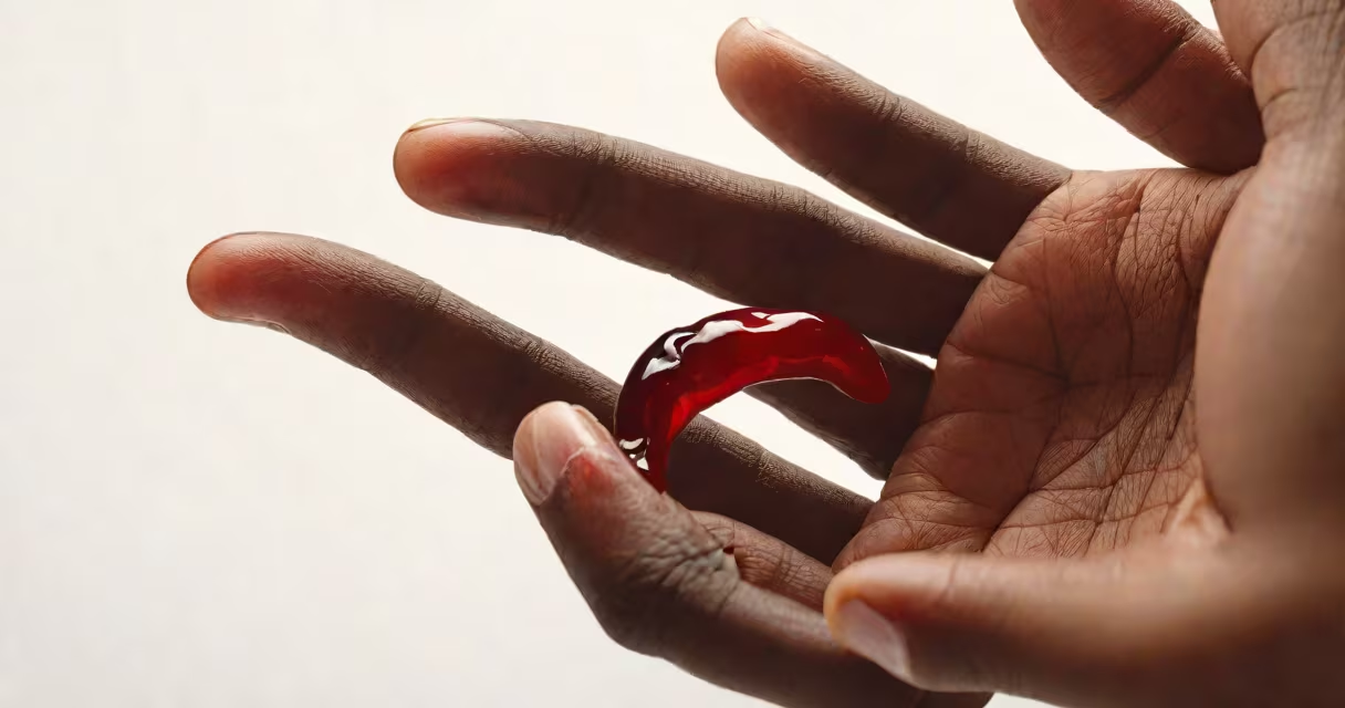
Sickle cell disease is a group of inherited blood disorders caused by a mutation in the hemoglobin gene, resulting in abnormally shaped red blood cells that resemble crescents or sickles. These misshapen cells obstruct blood flow, cause chronic anemia, and trigger episodes of severe pain. The condition manifests through visible physical signs, recurring health crises, and long-term complications affecting multiple organs.
The abnormal hemoglobin, known as hemoglobin S, replaces normal hemoglobin A. When oxygen levels drop, hemoglobin S polymerizes, distorting red blood cells into rigid, sticky sickle shapes. This deformation reduces oxygen delivery and damages tissues. The disease is autosomal recessive-both parents must carry the mutated gene for a child to inherit the full condition. It affects approximately 100,000 people in the United States and millions worldwide, primarily those of African, Mediterranean, Middle Eastern, and South Asian descent.
Visible signs of sickle cell disease often appear early in life. Infants may show jaundice due to rapid breakdown of sickled red blood cells. As children grow, they develop chronic pallor-pale skin and mucous membranes-from persistent anemia. This pallor results from low hemoglobin levels, typically below 8 g/dL in untreated cases.
Pain crises are among the most recognizable features. Episodes involve sudden, intense pain in bones, joints, abdomen, or chest. During a crisis, patients may appear distressed, clutching affected areas. The face may flush during acute episodes due to inflammation and vaso-occlusion. Swelling in hands and feet-known as dactylitis-is common in infants under two years old and presents as red, swollen digits that feel warm to touch.
Long-term effects include facial changes. Chronic anemia leads to bone expansion in the skull and facial bones, causing a characteristic "long face" appearance with prominent forehead and flattened nasal bridge. Some individuals develop leg ulcers near the ankles-open sores that resist healing due to poor circulation. These ulcers appear as shallow, irregular wounds with thickened edges and often drain serous fluid.
Blood tests reveal the sign of sickle cell disease: a high percentage of hemoglobin S. Newborn screening programs in the U.S. detect the condition within days of birth using high-performance liquid chromatography (HPLC) or capillary electrophoresis. A result showing >80% hemoglobin S confirms the diagnosis.
Imaging can reveal internal damage. An MRI of the brain may show silent cerebral infarcts-tiny strokes undetected by symptoms but visible as white matter lesions. Ultrasound of the spleen can identify splenic sequestration, where blood pools in the organ, causing sudden enlargement and shock. In adults, retinal exams may show hemorrhages or cotton wool spots indicating sickle cell retinopathy.
One non-obvious sign is the presence of "sickled" cells on a peripheral blood smear. Under a microscope, these cells appear as curved, elongated shapes resembling a crescent moon. They are fragile and prone to lysis, contributing to hemolytic anemia. However, not all sickled cells are visible in every sample-some only form under low oxygen conditions, which is why testing is often done under hypoxic conditions.
Sickle cell disease is frequently mistaken for other forms of anemia. Iron deficiency anemia causes small, pale red blood cells (microcytic hypochromic), while sickle cell anemia produces large, distorted cells with central pallor. Distinguishing them requires a hemoglobin electrophoresis test-not just a complete blood count.
Another misconception is that sickle cell disease only affects Black people. While it is most prevalent in populations of African ancestry, it also occurs in Hispanic, Indian, Arab, and Mediterranean communities. Over 1 in 365 Black American babies is born with sickle cell disease, but rates are rising in other groups due to migration and intermarriage.
A third error is assuming all sickle cell variants are equally severe. Hemoglobin SS (sickle cell anemia) is the most serious form. Hemoglobin SC disease causes milder symptoms but still leads to chronic pain and organ damage. Hemoglobin S-beta thalassemia varies widely in severity and may mimic either sickle cell or beta-thalassemia traits.
| Type | Hemoglobin Profile | Typical Severity | Common Symptoms |
|---|---|---|---|
| SS (sickle cell anemia) | 100% hemoglobin S | Severe | Pain crises, frequent infections, stroke risk |
| SC | 50% hemoglobin S, 50% hemoglobin C | Moderate | Chronic pain, joint issues, leg ulcers |
| S-beta thalassemia | Varying % hemoglobin S, reduced beta-globin | Mild to severe | Anemia, fatigue, jaundice |
In 2021, a 22-year-old man of Nigerian descent presented to the emergency department with severe left lower quadrant abdominal pain lasting six hours. He had a history of recurrent pain crises since age 5. On exam, he was pale, febrile (38.7°C), and had tenderness in the left lower quadrant. His hemoglobin was 7.8 g/dL, and a peripheral smear showed numerous sickle-shaped cells.
He was diagnosed with a vaso-occlusive crisis involving the mesenteric arteries. Imaging revealed bowel wall thickening consistent with ischemia. After IV hydration and morphine, his pain improved over four hours. He was admitted for observation and began hydroxyurea therapy. This case illustrates how sickle cell disease presents clinically: acute pain, anemia, and organ-specific ischemia triggered by sickled cells blocking microvasculature.
During my time working in a pediatric hematology clinic, I observed a 4-year-old girl with chronic leg ulcers. She had lived with sickle cell disease since birth. Her ulcers were resistant to standard wound care. We later discovered she had a rare variant with elevated hemoglobin S and impaired nitric oxide metabolism, which worsened tissue perfusion. This case reinforced that not all manifestations follow textbook patterns-and treatment must be individualized.
Sickle cell disease appears as a constellation of physical and medical signs: pallor, swelling in extremities, painful episodes, chronic anemia, and long-term organ damage. Visible indicators include jaundice, leg ulcers, and facial bone changes. Internally, it shows through abnormal blood smears, imaging findings, and lab results confirming hemoglobin S. Recognizing these signs early improves outcomes and guides timely intervention.