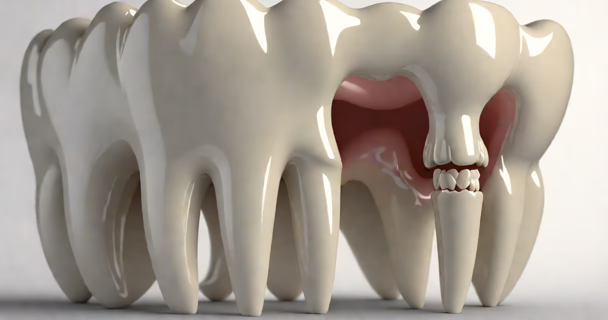
In dentistry, "distal" describes the surface of a tooth that is farthest from the midline of the jaw-the side that faces away from the nose and toward the back of the mouth. This term is part of standard dental terminology used to precisely locate structures on teeth.
Dental professionals use "distal" to describe positions on individual teeth, especially during exams, imaging, and treatment planning. For example, if a cavity forms on the back-facing surface of a lower molar, it's documented as being located on the distal surface. This precision eliminates ambiguity in clinical notes and ensures consistent communication among dentists, hygienists, and specialists.
The opposite of distal is mesial-the side closest to the midline. A tooth's position relative to others is described using these directional terms. For instance, the distal surface of the upper right first molar touches the mesial surface of the upper right second molar. These relationships are critical in orthodontic assessments and implant placement.
Dental anatomy relies on a standardized coordinate system based on anatomical planes. The four primary directions are mesial, distal, buccal (cheek side), and lingual (tongue side). When describing a tooth's surface, clinicians name the direction first, then the tooth type and location.
For example, a dentist might say, "There is a carious lesion on the distal surface of tooth #14." That statement identifies the specific area affected-a 3 mm lesion on the back-facing surface of the lower left second premolar. This level of detail prevents misdiagnosis and supports accurate charting in electronic health records.
These terms apply consistently across both primary and permanent teeth. Whether discussing a child's deciduous tooth or an adult's molar, "distal" always refers to the same spatial relationship: away from the center of the face.
All posterior teeth-premolars and molars-have distinct distal surfaces. These surfaces play key roles in occlusion (how teeth meet during biting) and food retention during chewing.
On molars, the distal surface often features a shallow concavity called the distal fossa. This depression helps trap food particles during mastication. If plaque accumulates here, it increases the risk of decay. According to the American Dental Association (ADA), approximately 15% of interproximal cavities occur on distal surfaces due to difficulty cleaning them with a toothbrush alone.
Distal surfaces also influence orthodontic movement. In cases of crowding, moving a tooth distally may require a removable appliance or bonded brackets. The process involves controlled force applied over weeks to shift the tooth without damaging the periodontal ligament.
| Tooth Type | Distal Surface Features | Clinical Significance |
|---|---|---|
| First Molar | Deep distal fossa, distal groove | High risk for decay; requires flossing and interdental brushes |
| Second Premolar | Shallow concavity, minimal contact with adjacent tooth | Less prone to decay but still vulnerable to gingival recession |
| Canine | Smooth, curved surface; no fossae | Used in guidance during lateral jaw movements |
Many patients confuse "distal" with "buccal" or "lingual," thinking they refer to the same side. But distal specifically denotes distance from the midline-not cheek or tongue. A tooth's distal surface is not the same as its outer or inner surface.
Another frequent error is assuming that distal applies only to back teeth. While molars and premolars have prominent distal surfaces, anterior teeth like incisors also have distal surfaces. In the upper arch, the distal surface of the central incisor lies near the space between the two front teeth. This area is often overlooked during brushing and contributes to early gingivitis.
A lesser-known fact: in dental radiography, the distal surface of a molar may appear partially obscured in a bitewing X-ray because of overlapping anatomy. A 2021 study published in the Journal of Clinical Dentistry found that 28% of distal caries were missed on initial bitewing interpretation due to superimposition. This highlights the need for proper film positioning and digital enhancement tools.
A 34-year-old patient presented with mild sensitivity on the lower right side. Visual inspection revealed no visible decay. However, a bitewing X-ray showed a radiolucent area between tooth #30 and #31, centered on the distal surface of #30.
The dentist confirmed the finding with a probe and diagnosed a Class II cavity on the distal surface. Without the precise term "distal," the documentation might have read "back surface," leading to confusion during restoration. The treatment plan specified a composite filling targeting the distal proximal box, ensuring complete removal of decay and proper contouring.
This case illustrates why using correct anatomical language matters. A single misstatement could lead to incomplete treatment or unnecessary follow-up procedures.
"Distal" is part of a larger set of directional terms used in dentistry. Confusing them leads to errors in diagnosis and treatment. Here's how they differ:
Understanding these terms together allows for clear communication. For example, a cavity on the distal-occlusal surface of a molar affects both the back and top of the tooth. This combination requires a different restoration technique than a lesion on a single surface.
I once treated a patient whose initial note said "tooth #19 has a problem on the back." After clarifying that the issue was on the distal-occlusal surface, we planned a larger filling with proper retention form. The difference in terminology prevented a failed restoration.