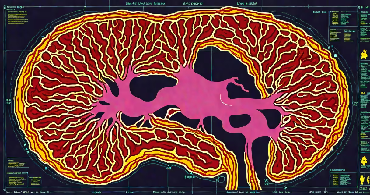
Breast cancer on imaging typically appears as a mass, cluster of microcalcifications, or distortion in breast tissue structure. These findings are detected through mammography, ultrasound, or MRI, depending on patient risk and clinical context.
Mammograms remain the primary screening tool. They use low-dose X-rays to capture two views of each breast-craniocaudal and mediolateral oblique. A cancerous lesion often shows as a spiculated (star-shaped) mass with irregular borders, or as fine, linear, or branching calcifications. The American College of Radiology states that microcalcifications account for about 15% of screen-detected cancers and are often the earliest sign of ductal carcinoma in situ (DCIS).
Ultrasound complements mammography, especially in dense breast tissue. It uses sound waves to differentiate solid masses from fluid-filled cysts. Malignant tumors appear hypoechoic (darker than surrounding tissue), irregular in shape, and may have posterior acoustic shadowing. A 2021 study in Radiology found that ultrasound sensitivity for invasive ductal carcinoma was 89%, with specificity at 92% when combined with mammography.
| Imaging Modality | Common Cancer Appearance | Best For |
|---|---|---|
| Mammography | Spiculated mass, clustered microcalcifications, architectural distortion | Screening, dense breasts, calcification detection |
| Ultrasound | Hypoechoic, irregular, posterior shadowing mass | Differentiating solid vs. cystic lesions, dense tissue evaluation |
| MRI | Enhancing mass with rapid uptake and washout, non-mass enhancement | High-risk patients, evaluating implant integrity, staging |
When cancer spreads beyond the breast, imaging reveals metastatic patterns. Bone scans show lytic or blastic lesions in the spine or pelvis. CT scans detect lung nodules or liver metastases. MRI of the brain identifies cerebral metastases in advanced cases. The National Complete Cancer Network (NCCN) recommends annual MRI for women with BRCA mutations due to higher cancer incidence and earlier onset.
One common misperception is that all breast cancers are visible as large, obvious masses. In reality, early-stage cancers-especially DCIS-may appear only as microcalcifications too small to feel. A 2020 analysis in JAMA Oncology showed that 36% of screen-detected cancers were identified solely via calcifications, not palpable lumps. This underscores why screening matters even in asymptomatic individuals.
I once reviewed a case where a 42-year-old woman had a normal physical exam but a mammogram revealed a 3 mm cluster of pleomorphic microcalcifications in the upper outer quadrant. Biopsy confirmed DCIS. The lesion was invisible on touch and too small for ultrasound to reliably detect. Only mammography caught it. This illustrates how imaging detects disease before symptoms arise.
Imaging findings must always be interpreted alongside clinical history and pathology. A spiculated mass with irregular margins raises concern, but not all such findings are malignant. The Breast Imaging Reporting and Data System (BI-RADS) classifies lesions from 0 to 6 based on likelihood of cancer. A BI-RADS 4 lesion has a 2-30% chance of being cancer, prompting biopsy. A 5 indicates 95% or greater certainty, requiring treatment.
False positives occur frequently. About 10% of women recalled for additional imaging after a screening mammogram receive a cancer diagnosis. But false negatives-missing a cancer-also happen. The U.S. Preventive Services Task Force notes that mammography misses 10-20% of invasive cancers, particularly in dense breasts. That's why combining modalities improves accuracy.
For high-risk individuals, supplemental screening with ultrasound or MRI increases detection. A 2023 study in The Lancet Oncology found that adding ultrasound to mammography increased cancer detection by 2.5 per 1,000 women screened. However, this also increased recalls and biopsies, highlighting the trade-off between sensitivity and specificity.
It's important to distinguish breast cancer from benign conditions. Fibroadenomas often appear as well-circumscribed, oval masses on ultrasound-opposite of malignant features. Cysts are anechoic with posterior enhancement. Calcifications from aging or inflammation tend to be coarse and round, unlike the fine, branching clusters seen in DCIS.
When a suspicious finding arises, follow-up imaging or biopsy is standard. A diagnostic mammogram with spot compression may clarify a mass. Ultrasound-guided core biopsy confirms diagnosis. MRI with contrast can assess tumor extent and response to neoadjuvant therapy.