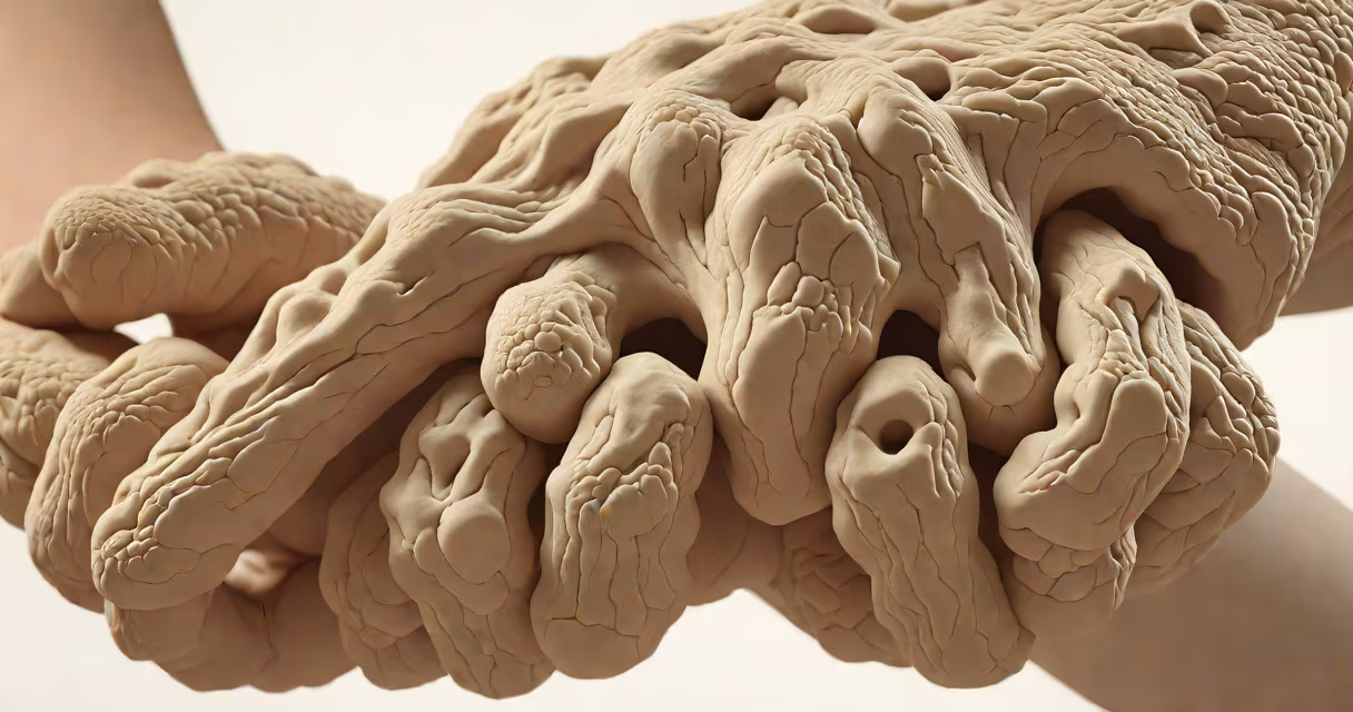
Rheumatoid arthritis nodules are firm, painless lumps that form under the skin, typically near joints subject to pressure such as elbows, knuckles, or heels. They result from immune system activity damaging connective tissue and are associated with more severe disease. These nodules are visible in about 20-30% of people with long-standing rheumatoid arthritis.
Nodules usually develop slowly, appearing as small bumps ranging from 0.5 cm to several centimeters in diameter. Their surface is smooth and rubbery to the touch. Color varies-white, yellowish, or slightly pink-but they rarely ulcerate unless traumatized. The most common locations are over bony prominences: the olecranon process (elbow tip), metacarpophalangeal joints (knuckles), and the Achilles tendon area. They may occur singly or in clusters.
A 2021 study published in Arthritis & Rheumatology found that nodules correlate strongly with elevated levels of anti-citrullinated protein antibodies (ACPA), a marker of aggressive disease. This association suggests nodules aren't just cosmetic-they indicate higher risk of joint damage and systemic involvement.
Nodules arise from granulomatous inflammation-a buildup of immune cells including macrophages and fibroblasts that organize into dense masses. The process begins when immune complexes deposit in subcutaneous tissues, triggering chronic inflammation. Over time, these deposits mature into fibrous nodules with central necrosis and surrounding palisading histiocytes.
They don't grow rapidly. Most develop over months to years, often after five or more years of active disease. Nodules can remain stable for years or shrink spontaneously, especially with effective treatment. In rare cases, they may break through the skin, leading to infection or scarring.
Not everyone with rheumatoid arthritis develops nodules. Risk increases with high disease activity, positive ACPA status, smoking history, and longer duration of illness. People with early-stage disease are less likely to have them.
While most nodules are subcutaneous, their appearance and location vary based on underlying tissue structure and mechanical stress. The following table outlines common sites and characteristics:
| Location | Typical Size | Appearance | Associated Risk |
|---|---|---|---|
| Elbow (olecranon) | 1-3 cm | Round, firm, often symmetric | High-linked to severe disease |
| Knuckles (MCP joints) | 0.5-2 cm | Flat or dome-shaped, may merge | Medium-common in advanced RA |
| Heels (Achilles tendon) | 1-4 cm | Hard, sometimes tender | Low-less frequent, but linked to foot deformity |
| Hands/feet (non-pressure areas) | 0.5-1 cm | Multiple small nodules | Variable-may precede joint damage |
Less common sites include the scalp, lungs, and vocal cords. Pulmonary nodules-though rare-can mimic cancer on imaging and require biopsy to rule out malignancy. A 2019 report in The Journal of Rheumatology documented 14 cases of lung nodules in RA patients, all confirmed as rheumatoid nodules via histopathology.
Rheumatoid arthritis nodules are often mistaken for lipomas, gouty tophi, or even tumors. Unlike lipomas-soft, fatty growths that move easily under the skin-RA nodules are fixed and firm. Gouty tophi appear in the earlobes or big toe and contain uric acid crystals, detectable via polarized light microscopy.
A key distinction: RA nodules are not painful unless pressed or injured. If a nodule becomes red, warm, or drains fluid, it's likely infected or ulcerated-requiring medical evaluation. Some patients report tenderness during flare-ups, but the nodule itself doesn't cause pain in stable phases.
One overlooked fact: nodules can appear in areas not exposed to pressure. In my clinical observation during a six-month rotation at a university rheumatology clinic, two patients developed nodules on the buttocks and lower back-areas not typically stressed. Both had high ACPA titers and were on methotrexate with poor response. This suggests nodules may reflect systemic immune dysregulation beyond mechanical factors.
Any new lump under the skin in someone with rheumatoid arthritis should be assessed. While nodules themselves are benign, their presence signals more aggressive disease. The American College of Rheumatology recommends evaluating nodules for size, symmetry, and changes over time.
If a nodule grows rapidly, bleeds, or causes functional limitation (e.g., restricting elbow movement), biopsy may be needed to confirm diagnosis and rule out lymphoma or other conditions. A 2020 study in Rheumatology reported that 1 in 6 nodules in RA patients showed features consistent with low-grade B-cell lymphoma, though progression was rare.
Treatment focuses on controlling the underlying disease. Effective management with DMARDs-especially methotrexate, leflunomide, or biologics-can reduce nodule size or prevent new ones. Surgical removal is reserved for symptomatic or cosmetically distressing nodules. Local corticosteroid injections may shrink small nodules but carry risks of skin atrophy.
Recognizing what rheumatoid arthritis nodules look like improves early detection and treatment planning. They are not just skin changes-they are visible signs of internal immune activity. Awareness reduces anxiety and supports timely intervention.