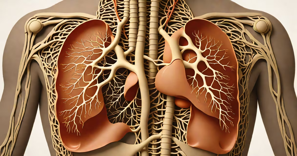
The respiratory system is the network of organs and tissues responsible for bringing oxygen into the body and expelling carbon dioxide through breathing. It includes the nose, pharynx, larynx, trachea, bronchi, bronchioles, alveoli, and the diaphragm. This system enables gas exchange between air and blood, supporting cellular respiration.
When you inhale, air travels through the nasal passages or mouth, down the pharynx and larynx, through the trachea, and into the bronchial tree. The trachea splits into two primary bronchi-one leading to each lung. Inside the lungs, the bronchi divide into smaller bronchioles, ending in clusters of tiny air sacs called alveoli. These alveoli are surrounded by capillaries, where oxygen diffuses into the bloodstream and carbon dioxide moves out.
Exhalation reverses the flow. The diaphragm contracts during inhalation, expanding the chest cavity and lowering pressure in the lungs. During exhalation, the diaphragm relaxes, reducing volume and pushing air out. This process happens about 12 to 20 times per minute at rest, adjusting based on activity level.
Each breath delivers approximately 500 milliliters of air to the lungs. Of that, about 150 mL remains in the conducting airways-the dead space-and never reaches the alveoli. Only the remaining 350 mL enters the gas-exchange zones. In the alveoli, oxygen crosses the thin epithelial barrier into red blood cells, binding to hemoglobin. Carbon dioxide, a metabolic waste product, follows the reverse path.
Alveolar surface area totals about 70 square meters-roughly the size of a tennis court-maximizing efficiency. The alveolar membrane is less than 0.5 micrometers thick, allowing rapid diffusion. A single alveolus measures about 200 micrometers in diameter, and each lung contains roughly 300 million alveoli.
Without this system, cells would lack oxygen within minutes. Brain function begins to fail after just 4 to 6 minutes without oxygen. The respiratory system maintains blood pH by regulating CO₂ levels-too much CO₂ causes acidosis, too little leads to alkalosis.
The respiratory system consists of upper and lower airways, lungs, and accessory muscles. The upper airway includes the nose, nasal cavity, pharynx, and larynx. The nose filters, warms, and humidifies incoming air. The nasal mucosa contains cilia and mucus to trap particles larger than 10 micrometers.
The lower airway comprises the trachea, bronchi, bronchioles, and alveoli. The trachea is reinforced with C-shaped cartilage rings to prevent collapse. Bronchi branch into smaller divisions until reaching terminal bronchioles, which lead to respiratory bronchioles and then alveolar ducts.
| Component | Function | Size/Detail |
|---|---|---|
| Nose | Filters, warms, humidifies air | 100,000+ cilia per square centimeter |
| Trachea | Conducts air to bronchi | 1.5 cm wide, 11-12 rings of cartilage |
| Alveoli | Site of gas exchange | ~200 μm diameter, 300 million per lung |
| Diaphragm | Primary muscle of breathing | Contracts to increase thoracic volume |
During exercise, breathing rate increases from 12 to 40 breaths per minute. Minute ventilation rises from 6 L/min at rest to over 100 L/min during maximal effort. The respiratory center in the medulla oblongata controls this automatic response based on blood CO₂ levels, not oxygen.
Many assume the lungs are involved in producing oxygen. They do not. The lungs only help its transfer from air to blood. Oxygen originates in photosynthesis by plants and algae. Humans rely entirely on atmospheric oxygen, which makes up 21% of dry air.
Another error is thinking that deep breathing increases oxygen saturation. While it improves ventilation, healthy individuals already saturate blood at 98% or higher at sea level. Hyperventilation reduces CO₂, causing dizziness and tingling due to respiratory alkalosis-not increased oxygen.
A lesser-known fact: the respiratory system shares embryological origin with the digestive tract. Both develop from the endoderm layer of the embryo. The trachea forms from a diverticulum of the primitive foregut around week 4 of gestation. This explains why some people have tracheoesophageal fistulas-abnormal connections between the airway and esophagus.
Asthma illustrates how dysfunction in the respiratory system disrupts normal function. In asthma, immune cells release histamine and leukotrienes, causing bronchoconstriction and inflammation. Airways narrow, increasing resistance to airflow.
During an attack, peak expiratory flow rate (PEFR) drops below 70% of predicted values. A person may breathe rapidly (tachypnea), use accessory muscles, and exhibit wheezing-a high-pitched sound caused by turbulent airflow through narrowed airways. Spirometry testing shows reduced forced expiratory volume in one second (FEV₁) and FEV₁/FVC ratio.
Treatment involves inhaled corticosteroids to reduce inflammation and short-acting beta-agonists like albuterol to relax smooth muscle. In severe cases, oral steroids or biologics may be required. Without treatment, persistent obstruction can lead to chronic hypoxia and pulmonary hypertension.
I once observed a patient with severe asthma who had a PEFR of 25% of predicted. After nebulized albuterol, their flow rate rose to 75%. The improvement was immediate-within 10 minutes, they stopped using accessory muscles and spoke in full sentences. This demonstrated how reversible airway narrowing can be with targeted therapy.
The respiratory system is often confused with the circulatory system. While both deliver oxygen, the circulatory system transports it via blood vessels. The respiratory system brings oxygen in and removes CO₂. The two systems work together but are distinct in structure and function.
Similarly, the kidneys regulate pH by excreting hydrogen ions and reabsorbing bicarbonate. The respiratory system handles acute pH changes by altering breathing rate. Chronic acid-base imbalances involve both systems. Renal compensation takes hours to days; respiratory compensation acts within minutes.
According to the National Institutes of Health (NIH), the average adult male has a total lung capacity of about 6 liters. The important capacity-the maximum air a person can exhale after a full inhalation-is typically 4.5 liters. These values vary with age, sex, height, and health status.
The American Lung Association confirms that smoking damages cilia in the respiratory tract, impairing mucus clearance. Over time, this leads to chronic bronchitis and emphysema-components of COPD. Smoking cessation improves lung function even in long-term smokers, though recovery is incomplete after decades of use.
Understanding what a respiratory system is requires recognizing its role in gas exchange, its anatomical components, and its integration with other bodily systems. It is not a passive conduit but an active, adaptive system essential for survival.
Every breath you take relies on this precise coordination of air movement, diffusion, and regulation. The respiratory system ensures your cells receive oxygen and remove waste-without it, life as we know it would cease.