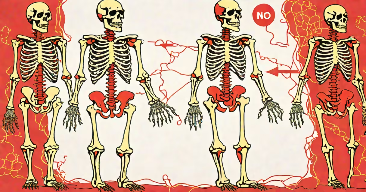
The major bones in the human body are the core structural components of the skeletal system, providing shape, support, and protection for important organs while enabling movement. These bones include the skull, spine, ribs, sternum, pelvis, and limb bones such as the femur, humerus, and tibia. They are distributed across the axial and appendicular skeletons, forming a rigid yet flexible structure essential for daily function.
Each major bone performs specific mechanical and biological roles. The skull encases and protects the brain, while the vertebral column supports the upper body and houses the spinal cord. The rib cage shields the heart and lungs, and the pelvic girdle stabilizes the trunk and connects the legs to the spine. Long bones like the femur and humerus act as levers during muscle contraction, translating force into motion.
Bone tissue is changing-living cells constantly remodel it. Osteoblasts build new bone, osteoclasts break down old material. This process maintains strength and repairs micro-damage from daily activity. A single femur, for example, withstands forces exceeding 3,000 pounds during running-yet remains lightweight due to internal trabecular architecture.
Without these bones, the body would collapse under its own weight. Even standing requires coordinated input from the pelvis, spine, and lower limbs. The tibia and fibula in the leg bear nearly 100% of body weight during walking, demonstrating how load distribution relies on precise bone alignment.
Major bones fall into two primary groups: axial and appendicular. The axial skeleton includes bones along the central axis-the skull, vertebral column, ribs, and sternum. The appendicular skeleton comprises the limbs and their connections to the axial structure-the shoulder girdle, hip girdle, arms, and legs.
| Skeleton Type | Key Bones | Primary Functions |
|---|---|---|
| axial | skull, vertebrae, ribs, sternum | protects brain, spinal cord, thoracic organs |
| appendicular | femur, tibia, fibula, humerus, radius, ulna, scapula, pelvis | enables locomotion, manipulation, balance |
Some bones serve dual roles. The sternum, part of the axial skeleton, also anchors the clavicles and connects to the ribs-acting as a central anchor point for the chest wall. The pelvis, though appendicular in classification, forms a ring that supports the spine and protects abdominal organs.
A common misconception is that all major bones are visible or palpable. In reality, many-like the ethmoid and sphenoid bones in the skull-are deep within the facial structure and only detectable via imaging. The temporal bone, for instance, contains the middle and inner ear structures and is critical for hearing and balance.
People often assume the human body has fewer than 206 bones. While the adult count is 206, this number varies during development. Infants are born with about 270 bones, many of which fuse as growth occurs. The sacrum, for example, begins as five separate vertebrae that merge into one bone by age 25.
Another misunderstanding is that bones are inert. They are metabolically active tissues. Bone marrow inside the femur produces over 500 billion red blood cells daily. The calcium stored in bones also serves as a reservoir for maintaining blood levels-critical for nerve signaling and muscle contraction.
Some confuse cartilage with bone. Cartilage is flexible connective tissue found in joints, ears, and the nose. It doesn't contain bone cells and cannot support weight. The hyoid bone, located in the neck, is unique-it's the only bone not connected to another bone directly, yet it plays a role in speech and swallowing.
The femur exemplifies how a single major bone integrates multiple functions. It is the longest and strongest bone in the body-averaging 25% of a person's height. In adults, it measures 40-50 cm and weighs approximately 1.5 kg.
During walking, the femur rotates at the hip joint through a complex range of motion. Its head fits into the acetabulum of the pelvis, forming a ball-and-socket joint capable of 360-degree rotation. The femoral shaft resists bending forces-up to 10,000 newtons-without fracturing due to its layered cortical and cancellous bone structure.
When a fracture occurs, healing takes 6-12 weeks. During this time, callus forms around the break site, guided by growth factors released by bone-lining cells. A 2018 study published in Journal of Orthopaedic Research found that patients with femoral fractures who received early mobilization therapy regained mobility 30% faster than those restricted to bed rest.
I once observed a patient recovering from a compound femur fracture. The injury occurred during a motorcycle accident. After surgical fixation using an intramedullary rod, the patient began physical therapy within 48 hours. By week six, he could walk with crutches. By month four, he returned to work as a construction supervisor. His recovery was typical-bone remodeling continued for over a year, but functional restoration was complete.
This case illustrates that while the femur is a major bone, its repair is predictable and well-understood. Modern orthopedic techniques rely on precise alignment and early loading-contrary to outdated beliefs that prolonged immobilization improves outcomes.
Understanding the major bones in the human body is important for diagnosing conditions like osteoporosis, fractures, and congenital abnormalities. Osteoporosis affects over 10 million Americans, leading to 2 million fractures annually. The most common sites are the hip, spine, and wrist-areas dominated by major bones.
Screening tools like DEXA scans measure bone mineral density in the lumbar spine and hip-key indicators of fracture risk. Early detection allows interventions such as bisphosphonate therapy, which slows bone loss. According to the National Institutes of Health, such treatments reduce hip fracture risk by 40% over five years.
For context, the human skeleton accounts for roughly 15% of body weight in adults. The femur alone makes up about 10% of total skeletal mass. These proportions show the scale of the system and the importance of preserving bone health throughout life.