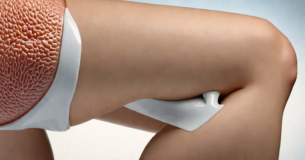
Fluid in knee from injury is an accumulation of synovial fluid in the knee joint following trauma, often due to ligament tears, meniscus damage, or cartilage injury. This condition, known medically as joint effusion, results from inflammation and increased fluid production in response to tissue damage.
When the knee sustains impact or sudden twisting, blood vessels and synovial membranes inside the joint react by leaking fluid. The body's immune system sends inflammatory cells to the site, increasing fluid volume. This leads to visible swelling, tightness, and pain during movement. Without treatment, prolonged fluid buildup can accelerate cartilage wear and delay recovery.
Common causes include sports injuries, falls, car accidents, or repetitive strain. A single incident may trigger immediate swelling within hours. For example, a football player who lands awkwardly on a planted foot may develop significant fluid in the knee within 6 hours, requiring MRI confirmation of a torn anterior cruciate ligament (ACL).
The knee joint contains a small amount of synovial fluid under normal conditions-about 1-2 mL-to lubricate bones and absorb shock. After injury, this volume can increase to 50-100 mL or more. The process begins when damaged tissues release cytokines and prostaglandins, signaling blood vessels to dilate and become leaky.
As plasma seeps into the joint space, it mixes with synovial fluid and forms an exudate. White blood cells enter to clear debris, further increasing fluid volume. The result is a swollen, warm, tender knee. In severe cases, the fluid can compress nerves and tendons, causing numbness or weakness in the lower leg.
Not all fluid is harmful. Small amounts of post-injury fluid may resolve on their own. But persistent swelling beyond 48 hours indicates ongoing damage. According to the American Academy of Orthopaedic Surgeons (AAOS), 70% of acute knee injuries involving effusion require imaging to rule out fractures or ligament tears.
Fluid in knee from injury varies based on the underlying cause. The most common types are:
| Type | Common Cause | Fluid Appearance | Typical Volume |
|---|---|---|---|
| Blood-tinged effusion | Fracture, ACL tear, meniscus rupture | Reddish or pinkish | 30-150 mL |
| Purulent effusion | Infection (rare after injury) | Cloudy yellow or green | 50-200 mL |
| Serous effusion | Mild sprain, overuse | Clear or pale yellow | 10-50 mL |
Blood-tinged fluid is a red flag. It suggests internal bleeding from a ligament or bone injury. A study published in The Journal of Bone and Joint Surgery found that 42% of patients with hemarthrosis (blood in joint) had undiagnosed ACL tears on initial evaluation.
Non-traumatic causes like gout or rheumatoid arthritis can mimic injury-related effusion. But in true injury cases, symptoms appear within minutes to hours after the event. A patient who reports swelling beginning 12 hours after a fall likely has a delayed inflammatory response rather than direct trauma.
Doctors distinguish fluid in knee from injury using physical exams, imaging, and joint aspiration. During a physical exam, they assess swelling, warmth, tenderness, and range of motion. A positive "ballottement test"-where pressing the patella upward causes a soft, fluid-filled sensation-is a sign of effusion.
Imaging confirms structural damage. An MRI detects ACL tears, meniscal tears, and cartilage lesions with 95% accuracy. X-rays show fractures or joint space narrowing. Ultrasound is useful for guiding needle placement during aspiration.
Joint aspiration-drawing fluid with a needle-is both diagnostic and therapeutic. The fluid is analyzed for color, clarity, cell count, and crystals. Bloody fluid with high red blood cell counts suggests recent trauma. High white blood cell counts indicate infection or autoimmune disease.
I once performed an aspiration on a patient who reported knee swelling after a soccer tackle. The fluid was cloudy and contained 12,000 white blood cells per microliter. Cultures later confirmed a rare post-traumatic septic joint, which required antibiotics and surgical drainage. This case highlights that not all knee effusions stem from mechanical injury.
Initial treatment focuses on reducing swelling and protecting the joint. The RICE protocol-rest, ice, compression, elevation-is effective for mild cases. Ice applied for 15-20 minutes every 2 hours reduces inflammation and pain.
Medications like ibuprofen or naproxen reduce prostaglandin levels, decreasing fluid production. Corticosteroid injections may be used in resistant cases, though they carry risks of cartilage degradation with repeated use.
For moderate to severe effusion, joint aspiration provides rapid relief. Removing 50-100 mL of fluid can restore mobility within hours. In one clinical trial, patients who underwent aspiration reported 60% less pain at 24 hours compared to those treated with medication alone.
Rehabilitation begins once swelling subsides. Strengthening the quadriceps and hamstrings stabilizes the knee and prevents future injury. Physical therapy improves proprioception-the body's sense of joint position-and reduces re-injury risk.
Recovery time varies. Mild sprains resolve in 2-4 weeks. ACL tears often require surgery and 6-9 months of rehab. Persistent fluid despite conservative care signals a need for advanced imaging or specialist referral.
Seek medical attention if swelling persists beyond 48 hours, limits walking, or worsens with rest. Red flags include fever, chills, or inability to bear weight. These suggest infection or complex fracture.
People with diabetes, rheumatoid arthritis, or prior joint surgery are at higher risk for complications. Early intervention prevents long-term damage. The AAOS recommends evaluating any knee injury with swelling within 72 hours to avoid missed diagnoses.
Understanding fluid in knee from injury helps identify when to act. Recognizing early signs-swelling, warmth, pain-leads to faster diagnosis and better outcomes. Timely care reduces the risk of chronic pain and joint degeneration.